Generative Deep Learning for Computational Destaining and Restaining of Unregistered Digital Pathology Images

Motivation
In clinical digital pathology, virtual tissue staining models are traditionally trained and evaluated using tightly paired, perfectly registered image datasets from a single institution. However, deploying these generative models in real-world clinical workflows introduces extreme domain shift due to variations in tissue processing, scanner hardware, and site-specific protocols. Furthermore, exact pixel-level registration between stained and unstained slices is rarely available in routine clinical practice. To translate AI-driven histopathology pipelines to scalable clinical settings, Dr. Pratik Shah’s lab evaluated whether preprocessing-based domain adaptation alone could recover cross-site model performance on entirely unseen, spatially unregistered external whole-slide image (WSI) datasets without any model retraining.
Technical Summary
This project demonstrates the cross-site robustness of pretrained conditional generative adversarial networks (cGANs) developed using the pix2pix framework with a U-Net generator and a PatchGAN discriminator. Models originally trained on 102 registered prostate core biopsy WSIs from Brigham and Women’s Hospital (dataset_B) were deployed directly on an external, out-of-distribution dataset of 82 spatially unregistered biopsy cores from Stanford University (dataset_S). To bridge the institutional domain shift without fine-tuning or modifying the model architecture, a dual-channel preprocessing pipeline was developed:
- H\&E Stained Images (GH\&E): Processed via Macenko stain normalization and global histogram alignment via cumulative distribution function matching against dataset_B reference frames.
- Ground-truth Unstained Tissue (GUS): Standardized using a custom channel-wise intensity calibration procedure with modified RGB channel weights to align color balance and saturation with the training distribution.
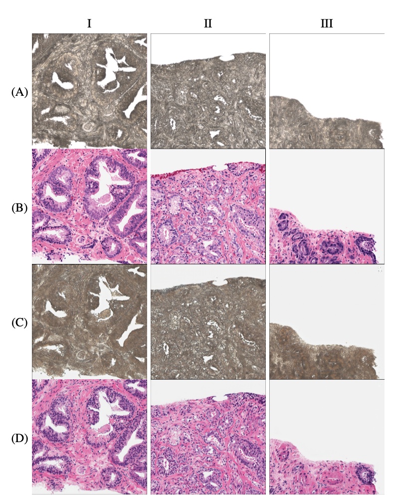
- Inference Pathways: Evaluated virtual destaining (GH\&E—>VDS), direct staining (GUS—> VH\&E), and an integrated digital destain-restain loop (GH\&E—>VDS —>VH\&ER). Because physical image registration was intentionally omitted to mirror actual clinical conditions, quantitative comparisons establish a conservative, real-world lower bound for generative deep learning performance in pathology.
FAQ
What is the value proposition of this work?
This research proves that previously trained generative models can successfully infer across different healthcare institutions by implementing smart, non-invasive input preprocessing rather than costly, resource-intensive model retraining. Additionally, it introduces an integrated “destain-restain digital loop” (GH\&E—>VDS—> VH\&ER) which serves as a vital indirect validation strategy for clinics where matching, paired ground-truth unstained physical tissue is unavailable.
What were the key findings from this work?
- Successful cross-site mapping: Under challenging, unregistered deployment conditions, virtual destaining achieved a solid Pearson correlation coefficient (PCC) of 0.854 ± 0.04, a structural similarity index measure (SSIM) of 0.699 ± 0.11, and a peak signal-to-noise ratio (PSNR) of 18.41 dB.
- Digital loop superiority: The two-stage destain-restain loop (VH\&ER vs. GH\&E) consistently outperformed direct computational staining from raw unstained inputs across all quantitative performance markers (PCC: $0.798 vs. 0.715; SSIM: 0.756 vs. 0.718; PSNR: 20.08 dB vs. 18.51 dB).
- Input harmonization bottleneck: The superiority of the digital loop implies that input harmonization and preprocessing quality represent a more critical real-world bottleneck to cross-site AI performance than the intrinsic capacity of the generative network.
- Direct staining limits: Pixel intensity profiling confirmed that direct staining models capture global contrast well, indicating that its main constraint in cross-site deployment is structural fidelity rather than color or brightness mismatch.
What are the main outcomes and the meaning of this work for deep learning research and clinical applications?
The study provides a reliable foundation for scaling virtual tissue processing in digital pathology without requiring institutions to gather massive, perfectly aligned paired training sets locally. By verifying model translation boundaries on external data, this architecture can safely guide automated downstream clinical tasks—such as computational H\&E virtual staining for rapid diagnosis, while preserving physical biopsies for subsequent molecular profiling and molecular assays.
What are the next steps?
Future growth involves expanding this cross-site validation paradigm across larger multi-institutional cohorts, different organ systems, and alternative stain modalities. Furthermore, research will focus on developing targeted domain-adaptation layers to explicitly protect complex clinical features during the virtual translation process.
What are the limitations and future growth areas of this research?
Assessment by a board-certified anatomic pathologist revealed a performance split between tissue compartments: benign structures (stroma, fat, and glandular architecture) were perfectly maintained, but malignant cancer glands occasionally suffered morphological degradation, sometimes rendering with vessel-like muscular wall profiles. Addressing this localized geometric breakdown in tumor architecture is a goal for future domain-adaptation iterations.
Publications
- Generative deep learning for computational destaining and restaining of unregistered digital pathology images. 2026. arXiv:2605.14251 preprint [https://doi.org/10.48550/arXiv.2605.14251].
Contributors and coauthors
Aarushi Kulkarni: Undergraduate student
Alarice Lowe: Clinical pathologist
Pratik Shah^: Principal investigator (^Senior and corresponding author).