Non-Ionizing Imaging for 3D Radiography instead of Cone-Beam Computed Tomography
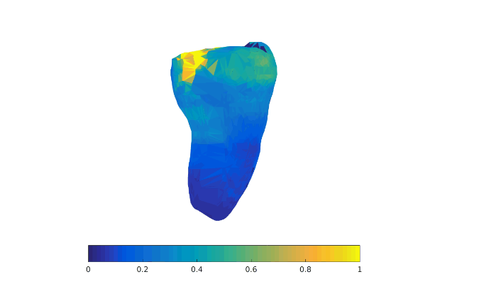
Three-dimensional radiographs are commonly used for evaluating sub-surface hard structures of teeth, but they have low sensitivity for early caries lesions, particularly those on tooth occlusal surfaces, and they are also frequently refused by patients over safety concerns. Our goal is to augment ultimately replace the prevalence of ionizing and expensive 3D X-ray imaging and cone-beam computed tomography (CBCT) for dental care with near-infrared (NIR) imaging. Translucency of teeth in the NIR range offers non-ionizing and safe detection of dental features. NIR can be used in conjunction with multiple light sources to create three-dimensional images of teeth. By modeling the scattering of photons in teeth, we can effectively see several millimeters inside, providing additional diagnostic value.
Why is this work important?
Three-dimensional radiographs and cone-beam computed tomography are commonly used for evaluating sub-surface hard structures of teeth. While radiographs are the current standard of care for diagnostic dental imaging, they have low sensitivity for early caries lesions, particularly those on tooth occlusal surfaces. They are also frequently refused by patients over safety concerns about exposure to ionizing radiation. Medical image acquisition without ionizing radiation can expand the use of important diagnostic tools and decrease safety concerns.
What has been done before?
NIR light can be transmitted across healthy dental enamel with marginal scattering, allowing for imaging dental features. NIR light at 850 nm and 1310 nm, which strike a balance between enamel and water attenuation, have been shown to provide helpful diagnostics that visual examination alone lacks. Our previous work has demonstrated the sensitivity of 850 nm NIR images to early caries lesions and demineralization. We think 3D NIR can synergistically augment or eventually replace ionizing radiation as the standard of care. We aim to expand 3D modelling of visible light and NIR imaging to tissue biopsy for rapid point-of-care imaging.
What are our contributions?
Preliminary work shows promise for augmenting the diagnostic power of NIR by modeling scattering of light.
What are the next steps?
Large-scale screenings can evaluate the effectiveness of our new imaging process.
Related projects
Dr. Pratik Shah
Faculty Member
Other Contributors
Aman Rana, Senior Research Support Associate
Gregory Yauney, Technical Associate
Keith Angelino, Technical Associate
Dr. David Edlund, DMD